Purple amethyst, the most prized variety of macrocrystalline quartz, forms through complex geological processes spanning millions of years in Earth’s crust. Understanding these formation mechanisms reveals why certain localities produce exceptional specimens while others yield only commercial-grade material. The interplay between temperature, pressure, chemical environment, and time creates the stunning purple crystals treasured throughout human history.
Amethyst crystallizes from silica-rich hydrothermal solutions in volcanic and metamorphic environments. The process begins when superheated water, enriched with dissolved silica (SiO₂) and trace elements, circulates through fractures and cavities in host rocks. As these solutions cool from temperatures exceeding 450°C to below 300°C, silica precipitates as quartz crystals. The critical factor distinguishing amethyst from colorless quartz is the presence of iron impurities and subsequent irradiation that creates the characteristic purple coloration.
The primary formation environments include volcanic geodes, pegmatite veins, and alpine-type fissures. Volcanic geodes, particularly abundant in Brazilian and Uruguayan basalt flows, form when gas bubbles in cooling lava create cavities subsequently lined with crystals. These geodal deposits produce the majority of commercial amethyst, with individual geodes ranging from fist-sized specimens to cathedral geodes exceeding 3 meters in height. The growth sequence typically begins with chalcedony or agate bands, followed by successive layers of quartz crystals growing toward the cavity center.
Pegmatite-hosted amethyst forms during the final crystallization stages of granitic intrusions. These environments produce exceptional clarity specimens, though typically smaller than geode crystals. The Four Peaks mine in Arizona exemplifies pegmatite-type deposits, yielding deep purple crystals highly valued for faceting. Alpine-type veins, formed by metamorphic processes in mountain-building events, produce the legendary specimens from localities like Vera Cruz, Mexico, characterized by exceptional transparency and distinctive crystal habits.
Crystallographic Structure and Properties
Amethyst exhibits trigonal crystal symmetry, belonging to crystal class 32 in the hexagonal crystal system. The fundamental unit cell contains three SiO₄ tetrahedra arranged in a helical pattern along the c-axis. This atomic arrangement creates either left-handed or right-handed spirals, determining the crystal’s optical rotation properties. The unit cell parameters measure a = 4.913 Å and c = 5.405 Å, with each silicon atom tetrahedrally coordinated to four oxygen atoms at Si-O distances of 1.605-1.611 Å.
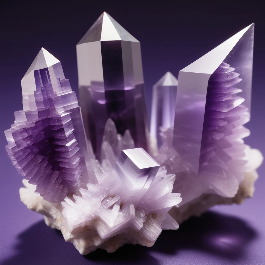
Crystal morphology varies significantly based on formation conditions. The most common form combines hexagonal prism faces {10-10} terminated by positive {10-11} and negative {01-11} rhombohedra. The ratio of these faces determines crystal habit, ranging from short, stubby crystals where rhombohedral faces dominate to elongated prismatic crystals in rapid-growth environments. Phantoms, visible growth zones within crystals, record changes in growth conditions over time, creating spectacular specimens prized by collectors.
Brazil law twinning, where two crystals intergrow at specific angles, occurs frequently in amethyst. This twinning follows a 180° rotation about the c-axis, creating penetration twins or contact twins. Japan law twins, involving 84°33′ rotation, produce distinctive V-shaped formations. These twinned crystals often display superior color distribution and interesting optical effects, making them particularly valuable for collectors and metaphysical practitioners.
Physical properties directly influence amethyst’s durability and workability:
- Hardness: 7 on Mohs scale (indentation hardness ~1,100 kg/mm²)
- Specific gravity: 2.65 (±0.01)
- Refractive indices: nω = 1.544, nε = 1.553
- Birefringence: 0.009
- Cleavage: None (conchoidal fracture)
- Thermal expansion: 13.4 × 10⁻⁶/°C perpendicular to c-axis
- Piezoelectric coefficient: d11 = 2.3 × 10⁻¹² C/N
- Optical activity: 21.7°/mm rotation at 589 nm
Color Origins and Mechanisms
The purple coloration in amethyst results from complex color centers involving iron impurities and natural irradiation. Iron substitutes for silicon in the crystal lattice, typically at concentrations of 10-100 parts per million. During formation, Fe³⁺ ions occupy tetrahedral sites, creating [FeO₄]⁰ centers. Natural gamma radiation from surrounding rocks transfers electrons, converting some Fe³⁺ to Fe⁴⁺, generating the characteristic O⁻-Fe⁴⁺ color centers responsible for purple coloration.
The relationship between iron concentration and color intensity follows a non-linear pattern. Optimal coloration occurs at 40-60 ppm iron, with higher concentrations producing grayish or brownish overtones. The irradiation dose determines color saturation, typically requiring 10⁶-10⁸ rads accumulated over geological time. This explains why amethyst from radioactive granite environments often displays more intense coloration than specimens from less radioactive basaltic hosts.
Color distribution patterns reveal growth dynamics and formation conditions. Sector zoning results from different growth rates on various crystal faces, creating alternating purple and colorless zones. Chevron amethyst displays distinctive V-shaped patterns from alternating growth of amethyst and white quartz layers. These patterns follow crystallographic orientations, with color concentrated along rhombohedral growth sectors while prismatic sectors remain colorless or lightly colored.
Temperature sensitivity of amethyst color centers has profound implications for treatment and care. Heating to 300-400°C begins breaking down color centers, causing gradual lightening. At 450-500°C, complete decolorization occurs, producing colorless quartz or, in iron-rich specimens, citrine through Fe³⁺ aggregation. This thermal instability necessitates careful temperature control during jewelry manufacturing and repair. Some deposits produce amethyst that turns green (prasiolite) upon heating, attributed to different iron site occupancies and intervalence charge transfer mechanisms.
Trace Element Chemistry and Geographic Signatures
Advanced analytical techniques reveal trace element signatures unique to specific amethyst deposits, enabling geographic origin determination. Laser ablation inductively coupled plasma mass spectrometry (LA-ICP-MS) detects elements at parts-per-billion concentrations, creating chemical fingerprints for provenance studies.
Brazilian amethyst typically contains aluminum (100-500 ppm), lithium (1-10 ppm), and germanium (0.1-2 ppm) substituting in tetrahedral sites. The Al/Li ratio often approaches 10:1, suggesting coupled substitution maintaining charge balance. Uranium and thorium at ppb levels provide natural radiation for color center formation. Rio Grande do Sul specimens show elevated manganese (5-20 ppm), contributing subtle red flashes in certain lighting conditions.
Zambian amethyst exhibits distinct chemistry with higher iron content (60-150 ppm) and significant titanium (10-30 ppm). This composition produces deep purple coloration with red and blue flashes, highly prized in fine jewelry. Calcium and magnesium at 50-200 ppm reflect the metamorphic host rock environment. The presence of rare earth elements, particularly cerium and lanthanum, distinguishes African material from South American sources.
Siberian amethyst, historically considered the finest quality, contains unique trace element assemblages including elevated germanium (2-5 ppm) and gallium (0.5-2 ppm). These elements potentially influence color center formation, contributing to the legendary “Siberian” deep purple with red flashes. Modern spectroscopic analysis reveals that historical “Siberian” color grades occur worldwide, though the term persists in quality description.
Spectroscopic Characteristics
Understanding amethyst’s interaction with electromagnetic radiation enables identification, quality assessment, and treatment detection. Various spectroscopic techniques reveal structural and chemical information invisible to conventional observation.
Ultraviolet-visible absorption spectroscopy shows characteristic bands at 545 nm and 520-530 nm from Fe⁴⁺ color centers, with intensity proportional to purple saturation. Additional weak bands at 380 nm and 950 nm arise from Fe³⁺ in different coordination environments. The ratio of these absorptions indicates heating history, as thermal treatment alters relative peak intensities. Prasiolite shows distinct absorption at 420 nm and 620 nm from Fe²⁺-Fe³⁺ intervalence charge transfer.
Infrared spectroscopy reveals hydroxyl groups and molecular water inclusions affecting clarity and stability. The fundamental OH stretching region (3000-3700 cm⁻¹) contains multiple bands indicating different hydrogen environments. Sharp bands at 3585 cm⁻¹ and 3543 cm⁻¹ indicate AlOH defects, while broad bands around 3400 cm⁻¹ suggest molecular water in structural channels. These features help distinguish natural from synthetic amethyst, as synthetic material typically shows different hydroxyl signatures.
Raman spectroscopy provides non-destructive phase identification and stress analysis. The primary quartz bands at 464 cm⁻¹ (α-quartz) shift with applied stress, enabling strain mapping in mounted stones. Additional bands at 206, 265, 355, and 402 cm⁻¹ confirm α-quartz structure. Peak width analysis reveals crystallinity, with natural amethyst showing narrower peaks than synthetic material due to slower growth rates and better atomic ordering.
Photoluminescence spectroscopy under laser excitation reveals trace element activators and defect centers. Green laser (532 nm) excitation produces red emission from Fe³⁺ centers, while UV excitation (365 nm) generates blue emission from Al-related defects. Time-resolved measurements distinguish different luminescence centers, providing detailed information about defect structure and formation conditions. These techniques prove invaluable for detecting treatments and enhancements.